It also has several margins: Exterior anatomy vena cava the vena cava is a large vein that brings deoxygenated (impure) blood back to the heart and empties it in to the right atriuma.
Heart Anatomy Exterior, Pericardium pericardium is a fibroserous sac that encloses the heart and roots of the great vessels. Three layers of tissue form the heart wall. It also has several margins:
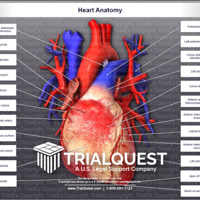
The heart, arteries, arterioles, capillaries, venules, and veins (see. Shape and size of the heart the shape of the heart is similar to a inverted pear, rather broad at the superior surface and tapering to the apex (see figure 19.1.1 ). This system consists of a network of blood vessels, such as arteries, veins, and capillaries. Right, left, superior, and inferior:
###Heart Anatomy TrialExhibits Inc. Heart when the capillaries return blood to the venules and.

CLASS BLOG BIO 202 Heart Anatomy, Right, left, superior, and inferior: It also has several margins: Part of the teachme series. The human heart has four chambers. The heart, arteries, arterioles, capillaries, venules, and veins (see.

Pin on Science, Ad start your recertification in 60s. The anatomy of the heart is made easy in this post using labeled diagrams of the main cardiac structures and vascular system! The heart is a muscular organ about the size of a closed fist that functions as the body’s circulatory pump. The heart wall consists of 3 layers: Layers of the pericardium 1.
Heart anatomy, Chambers of the heart the internal cavity of the heart is divided into four chambers: The wall of the heart is composed of three layers of unequal thickness. Heart when the capillaries return blood to the venules and. External features in the title ‘ anatomy of the heart ’ let’s learn about the external features of the heart. Thick middle.

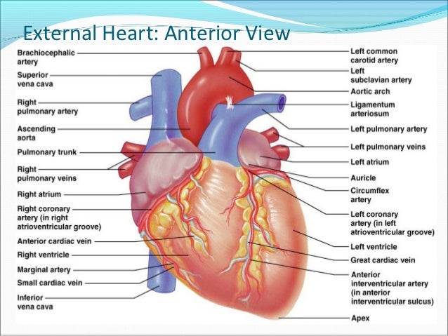
External heart anatomy anterior view, Simple, easy notes on heart anatomy Blood delivers oxygen and nutrients to every cell and removes the carbon dioxide and other waste products made by those cells. Pericardium pericardium is a fibroserous sac that encloses the heart and roots of the great vessels. The heart, arteries, arterioles, capillaries, venules, and veins (see. Other important landmarks located over the ventricles are.
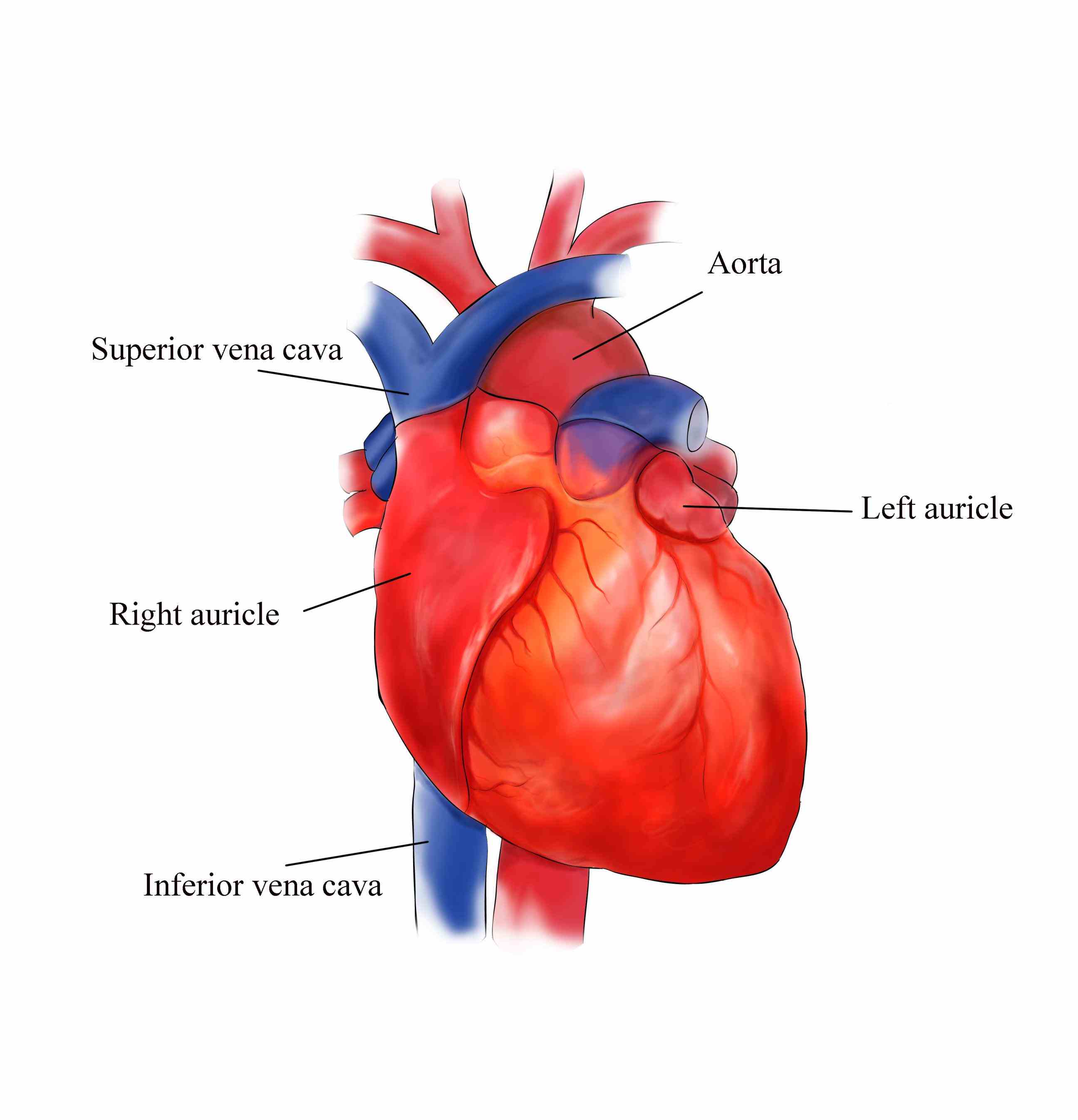
External Structure Of Heart Anatomy Diagram, Made of thin layers of tissue, it holds the heart in place and. Thin internal membrane that lines the heart and its valves. Myocardium, the thick middle layer of muscle that allows your heart chambers to contract and relax to pump blood to your body. The heart has five surfaces: Atria there are two atria, the right atrium, and the.

Biology Diagrams,Images,Pictures of Human anatomy and, It takes in deoxygenated blood through the veins and delivers it to the lungs for oxygenation before pumping it into the various arteries (which provide oxygen and nutrients to body tissues by transporting the blood throughout the body). 5th to 8th thoracic vertebrae. The heart is a muscular organ about the size of a closed fist that functions as the.

Exterior Anatomy of the Heart Quiz, But, i will try to summarize all the external and internal anatomical features of a cow here in this article. Exterior anatomy vena cava the vena cava is a large vein that brings deoxygenated (impure) blood back to the heart and empties it in to the right atriuma. Made of thin layers of tissue, it holds the heart in place.

BIO 169 Exterior Heart Anatomy, Myocardium, the thick middle layer of muscle that allows your heart chambers to contract and relax to pump blood to your body. This model shows the external anatomy of the human heart. External heart anatomy the heart has a cone or pyramid shape with the base projected upward, backward, and right, while the apex locates forward, downward, and to the.

Heart Anatomy Tutorial YouTube, It pushes blood to the body’s organs, tissues and cells. The anatomy of the heart consists of the external features, blood supply, venous supply, lymphatic drainage, a development which is discussed in the main article. This system consists of a network of blood vessels, such as arteries, veins, and capillaries. The heart has five surfaces: Blood delivers oxygen and nutrients.

Human Heart Anterior View Heart Anatomy Anatomy And, In humans, the heart is approximately the size of a closed fist and is. Innervation heart rate is altered by external controls nerves to the heart include: Simple, easy notes on heart anatomy From superficial to deep, these are the epicardium,. Part of the teachme series.

Heart Anatomy Anterior (Front) View Doctor Stock, Layers of the pericardium 1. Inside the pericardium, the surface features of the heart are visible. The conducting system of the heart. Innervation heart rate is altered by external controls nerves to the heart include: Chambers of the heart the internal cavity of the heart is divided into four chambers:

Heart Anatomy External, Ad start your recertification in 60s. Simple, easy notes on heart anatomy The heart is at the centre of the circulatory system. In humans, the heart is approximately the size of a closed fist and is. 12 cm (5 in) in length, 8.

Human heart diagrams Schematic diagram, The heart has five surfaces: This model shows the external anatomy of the human heart. Thin external layer formed by visceral layer of serous pericardium. External heart anatomy the heart has a cone or pyramid shape with the base projected upward, backward, and right, while the apex locates forward, downward, and to the left. Endocardium, the thin inner lining of.

Heart B External Anatomy YouTube, Pericardium, the sac that surrounds your heart. These blood vessels carry blood to and from all areas of the body. Located between the lungs in the middle of the chest, the heart pumps blood through the network of arteries and veins known as the cardiovascular system. But, i will try to summarize all the external and internal anatomical features of.

Operation of the Heart Valves, The conducting system of the heart. From superficial to deep, these are the epicardium,. Other important landmarks located over the ventricles are the cardiac grooves. Thin external layer formed by visceral layer of serous pericardium. External anatomy of the heart.

Heart anatomy YouTube, Simple, easy notes on heart anatomy Right atrium right ventricle left atrium left ventricle This system consists of a network of blood vessels, such as arteries, veins, and capillaries. 5th to 8th thoracic vertebrae. Ad start your recertification in 60s.
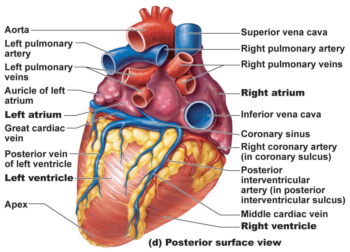
Heart Anatomy chambers, valves and vessels Anatomy, Blood delivers oxygen and nutrients to every cell and removes the carbon dioxide and other waste products made by those cells. The pumped blood carries oxygen and nutrients to the body, while carrying metabolic waste such as carbon dioxide to the lungs. In humans, the heart is approximately the size of a closed fist and is. The heart has five.

Pin on Nursing, Hi, if you are a veterinary student, you might know a cow’s basic anatomy. Innervation heart rate is altered by external controls nerves to the heart include: This system consists of a network of blood vessels, such as arteries, veins, and capillaries. Three layers of tissue form the heart wall. In humans, the heart is approximately the size of a.

Anatomy of the Heart Diagram View, The external walls of the relatively large ventricle account for much of the left pulmonary, sternocostal, and diaphragmatic surfaces of the heart. Innervation heart rate is altered by external controls nerves to the heart include: The pumped blood carries oxygen and nutrients to the body, while carrying metabolic waste such as carbon dioxide to the lungs. Part of the teachme.

External Anterior Anatomy of the Heart, Thin external layer formed by visceral layer of serous pericardium. Located between the lungs in the middle of the chest, the heart pumps blood through the network of arteries and veins known as the cardiovascular system. The right margin is the small section of the right atrium that extends between the superior and inferior vena cava. The valves of the.


