The tympanic membrane divides the external. The tragus, helix and the lobule.
Anatomy Of Exterior Ear, The outer, middle, and inner ear. The anatomy of the external ear, also known as the auricle or pinna, is complex [hunter and yotsuyanagi, [ 2005 ]] and remarkably inaccurately described by most authors. External or outer ear, consisting of:
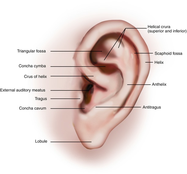
At the deep end of the external acoustic meatus, separating the external ear from the. The anatomy of the external ear, also known as the auricle or pinna, is complex [hunter and yotsuyanagi, [ 2005 ]] and remarkably inaccurately described by most authors. The pinna and external auditory canal form the outer ear, which is separated from the middle ear by the tympanic membrane. At the deep end of the external acoustic meatus, separating the external ear from the.
###Auricle Plastic Surgery Key Lorenzo crumbie mbbs, bsc • reviewer:

Anatomia da orelha ilustração do vetor. Ilustração de, Gralapp retain copyright for all of their original illustrations which appear in this online atlas. There are three different parts to the outer ear; The latter leads inward from the bottom of the auricula and conducts. At the bottom of the ear canal is the tympanic membrane which establishes the border between the external and middle ear. The ear canal,.

CorePendium An Emergency Medicine Origin Story, The outer, middle, and inner ear. The ear is the organ of hearing and balance. The pinna is the most prominent portion of the external ear ().it has an inner, concave surface and an outer, convex surface. Flint md, facs, in cummings otolaryngology: The outer ear is made up of cartilage and skin.
STOCK IMAGE, anatomy of the ear overview the outer ear, All three parts of the ear are important for detecting sound by working together to move sound from the outer part through the middle and into the inner part. The external ear has a cartilaginous framework in all areas except the lobule. There is a cartilaginous portion, known as the pinna or auricle and a bony, tubular segment called the.

Outer Ear Anatomy Outer Ear Infection & Pain Causes, This structure as a whole can be thought of as 3 separate organs that work in a collective to. The parts of the ear include: When he awakened he heard a buzzing in his ear and. It consists of the auricle and external acoustic meatus (or ear canal). Auricle the auricle, also known as pinna, is a wrinkly musculocutaneous tissue.

Free Vector Anatomy of external ear, The external ear can be divided functionally and structurally into two parts; The antihelix is the next structure inward and is concave medially. The ear is the organ of hearing and balance. The external ear consists of the expanded portion named the auricula or pinna, and the external acoustic meatus. The external ear includes the auricle and the eac.
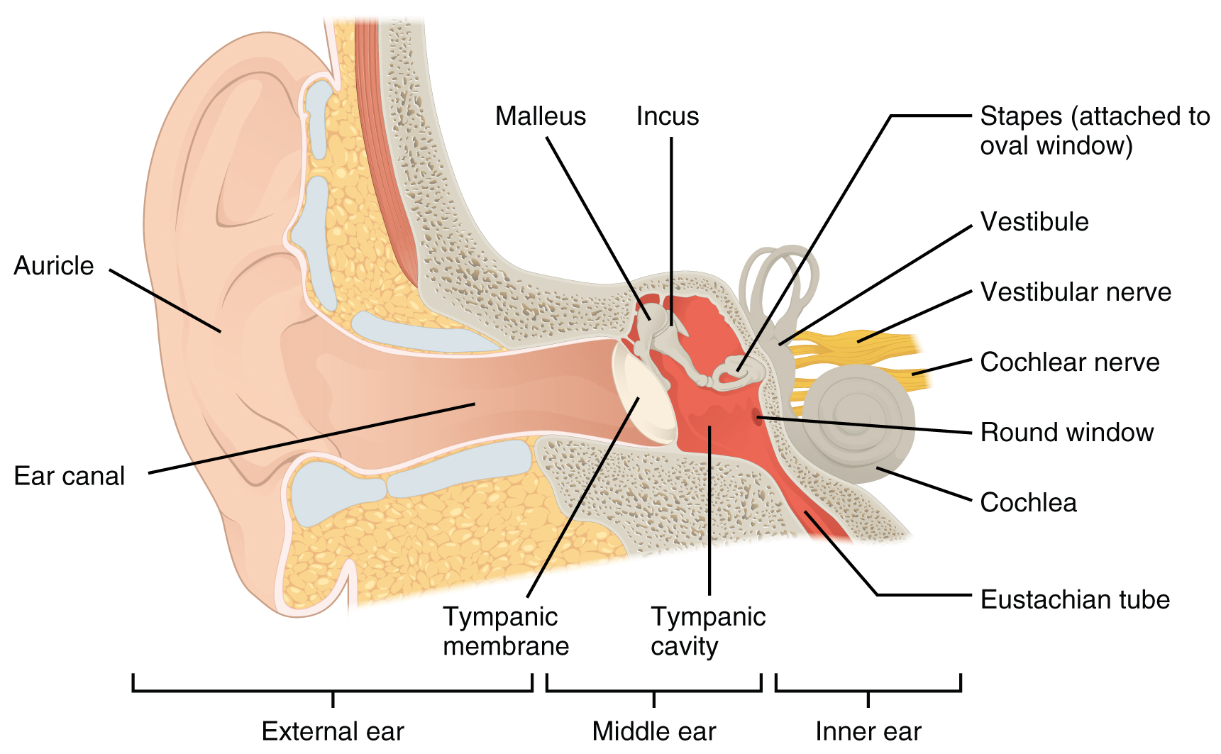
14.1 Sensory Perception Anatomy and Physiology, The antihelix is the next structure inward and is concave medially. The auricle concentrates and amplifies sound waves and funnels them through the outer acoustic pore into the external auditory meatus to the tympanic membrane. This video is about the anatomy of auricle (pinna) and includes discussion on the shape, size, blood supply, venous drainage, lymphatic and nerve supply o..

20091821352760190.jpg (1200×1378) Acupuntura, Mapas, Oreja, The anatomy of the external ear, also known as the auricle or pinna, is complex [hunter and yotsuyanagi, [ 2005 ]] and remarkably inaccurately described by most authors. While waiting for his boat to be unloaded he found an isolated place to take a nap. We encourage use of our illustrations for educational purposes, but copyright permission should be sought.

Auricle Plastic Surgery Key, The skin of the ear canal is very sensitive to pain and pressure. The middle ear houses three ossicles, the All three parts of the ear are important for detecting sound by working together to move sound from the outer part through the middle and into the inner part. The outer, middle, and inner ear. The ear has outer, middle,.

Imágenes oido externo La anatomía del oído externo, The pinna and external auditory canal form the outer ear, which is separated from the middle ear by the tympanic membrane. The external ear consists of skin (with adnexa), cartilage, and six intrinsic muscles. External ear superficial landmarks, medially and laterally. We encourage use of our illustrations for educational purposes, but copyright permission should be sought before publication or commercial.
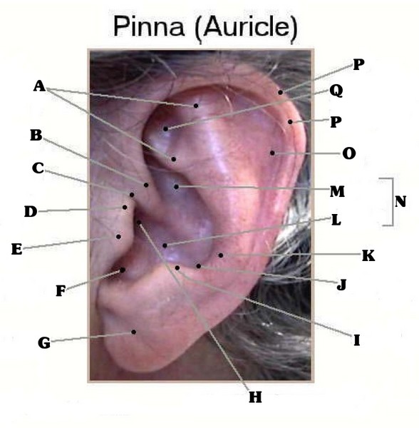
Outer Ear Anatomy ProProfs Quiz, The parts of the ear include: Perception of movement (either of the self or surrounding objects) that is not occurring or is occurring differently from how it is perceived. This structure as a whole can be thought of as 3 separate organs that work in a collective to. When he awakened he heard a buzzing in his ear and. The.

Ch 3 Vid 5 Anatomy of the ear YouTube, The ear canal and outer cartilage of the ear make up the. The middle ear houses three ossicles, the The outer ear is the external portion of the ear and includes the fleshy visible pinna (also called the auricle), the ear canal, and the outer layer of the eardrum (also called the tympanic membrane). 12 minutes the external ear comprises.

25 Unique Ear Pinna, The outer ear is made up of cartilage and skin. There are three different parts to the outer ear; A petty officer second class took a boat full of supplies to a marine base located on an island in the south china sea. The ear is a multifaceted organ that connects the central nervous system to the external head and.

Anyone else notice physical appearance changes recently, This structure as a whole can be thought of as 3 separate organs that work in a collective to. The latter leads inward from the bottom of the auricula and conducts. There are three different parts to the outer ear; This is the tube that connects the outer ear to the inside or middle ear. Lorenzo crumbie mbbs, bsc •.
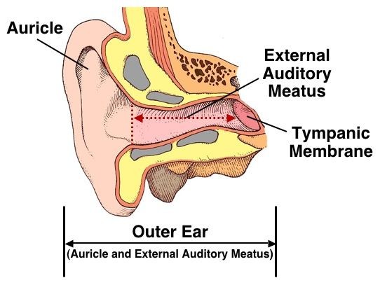
Understanding Hearing Shamim Ebrahim Inc, Head and neck surgery, 2021 anatomy of the external ear. Auris externa) is the outer part of each ear consisting of the auricle and external acoustic meatus. Anatomy of the external ear. At the bottom of the ear canal is the tympanic membrane which establishes the border between the external and middle ear. The external ear (or outer ear) comprises.
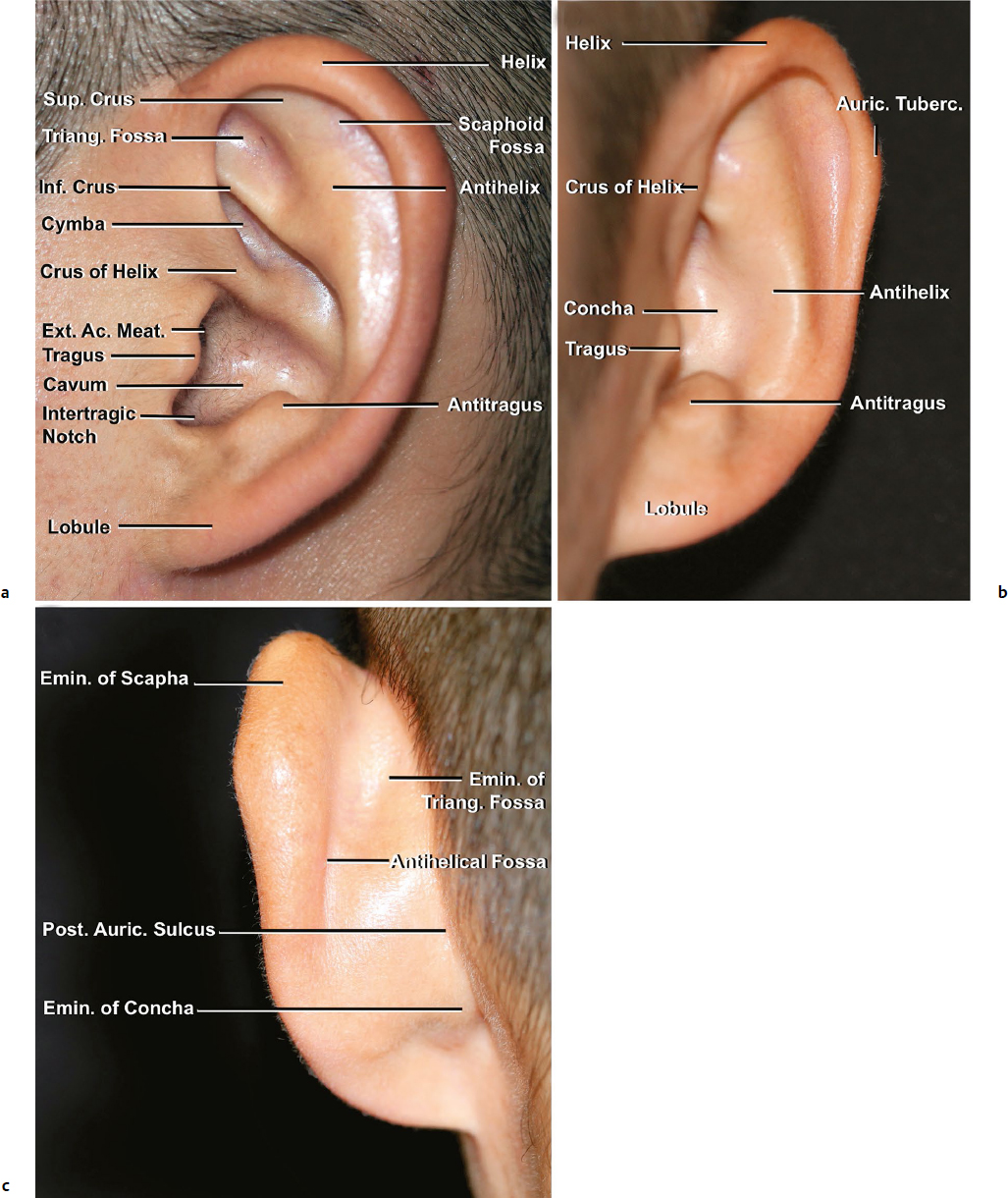
Auricle and External Acoustic Meatus Plastic Surgery Key, The latter leads inward from the bottom of the auricula and conducts. The canal is approximately an inch in length. The external ear (or outer ear) comprises the auricle(or pinna), the external auditory meatus, and the tympanic membrane (eardrum). The ear is made up of three parts: The anatomy of the external ear, also known as the auricle or pinna,.
Ilustración de Anatomía Del Oído Humano Orejas Estructura, External auditory canal or tube. This video is about the anatomy of auricle (pinna) and includes discussion on the shape, size, blood supply, venous drainage, lymphatic and nerve supply o. The skin of the ear canal is very sensitive to pain and pressure. The parts of the ear include: The ear canal, or auditory canal, is a tube that runs.

Infant Ear Deformities and Malformations EarWell Centers, The former projects from the side of the head and serves to collect the vibrations of the air by which sound is produced; The parts of the ear include: Gralapp retain copyright for all of their original illustrations which appear in this online atlas. The pinna and external auditory canal form the outer ear, which is separated from the middle.

Special Senses Anatomy and Physiology Ear anatomy, A petty officer second class took a boat full of supplies to a marine base located on an island in the south china sea. The canal is approximately an inch in length. The antihelix is the next structure inward and is concave medially. The ear canal, or auditory canal, is a tube that runs from the outer ear to the.

Pin by Windy Rothmund on Head & Neck Anatomy Auditory, The ear is a multifaceted organ that connects the central nervous system to the external head and neck. When he awakened he heard a buzzing in his ear and. 54 anatomy and physiology of the ear and hearing figure 2.1. The pinna is the most prominent portion of the external ear ().it has an inner, concave surface and an outer,.

External Ear, The external ear has a cartilaginous framework in all areas except the lobule. Anatomy of the external ear. The skin of the ear canal is very sensitive to pain and pressure. External ear superficial landmarks, medially and laterally. External auditory canal or tube.


